Development Of New Optical Techniques And Methods For Basic Biology And Biomedical Applications
The term “optical biopsy” identifies the diagnostic procedures based on the interaction between light and biological substrates, with their content in biomolecules acting as endogenous fluorophores when excited with light at a suitable wavelength.
The overall autofluorescence emission of cells, tissue and fluids depends on the kind, amount and microenvironment of the endogenous fluorophores, and their changes depending on alteration of normal state and rising of pathologies. As a consequence, autofluorescence analysis can provide diagnostic information, in real time and in the absence of external makers and dyes (label free).
The liver is engaged in several metabolic and catabolic activities to maintain the systemic homeostasis of nutrients and micronutrients, involving different endogenous fluorophores. These exhibit different distribution in the tissue and spectral properties, useful to appreciate both histological organization and composition of liver tissue, through suitable imaging or spectral analysis procedures.
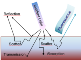 |
| The light interaction with a biological substrate results in various optical events, in including fluorescence. |

The contrast between the blue (NADH) and yellowish (lipofuscins) cytoplasmic areas and the light blue, brilliant reticular structures (fibrous proteins) allows to appreciate the plates of hepatocytes converging to the centrolobular vein (c) in normal parenchima, and the disorganized structure and the accumulation of collagen in the portal area (p) in fibrosis. In fatty liver (steatosis) lipid droplets are easily observable for their brilliant emission due to the presence o highly fluorescing vitamin A and fluorescing lipids.
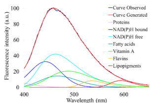
The different spectral properties of the liver fluorophores are identified by means of spectral function, which are used for the fitting analysis based estimation of the relative contribution of each fluorophore to the overall emission, similarly to an in situ biochemical analysis. Images and spectra are obtained under 366 nm excitation.
Cryostatic section of normal liver tissue
On these bases, autofluorescence analysis offers multiple diagnostic applications, from the real time monitoring of liver functionality in surgery and transplantation, to the histological diagnosis of liver biopsy. In the latter case, besides NAD(P)H and flavins, the emission signal of collagen and lipopigments is also used for the development of methods for the fast and cost effective screening of cases to avoid useless use of time in the selection of suspicious cases be sent to the pathologist for further observation, to confirm the diagnosis of liver disorder progression, from steatosis to most severe disease (hepatocarcinoma).
The increasing demand of new “serum panels” biomarkers for the non invasive diagnosis in hepatology and more generally in the study and therapy of systemic metabolic disorders has guided attention to the autofluorescence of serum. This is particularly promising because: i) liver is engaged in the storage and mobilization into the blood of retinol and lipids, in particular some free fatty acids, such as arachidonic, linoleic e oleic acids; ii) these compounds are fluorescing and can be estimated directly in crude serum; iii) retinol and some free fatty acids and their derivative (for example eicosanoids) have a pathophysiological meaning, since they act as antioxidants, regulators and mediators along the pathways of liver intrinsic or systemic regulation of defensive of harmful responses induced by hepatic stress.
 |
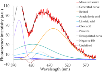 |
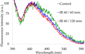
Emission spectra of serum collected from rats, controls vs hepatic ischemia and riperfusion (I/R), and an example of spectral fitting analysis (contro serum, fluorophres identified by colors). Fitting analysis allows to estimate the relative riising of arachidonic acid against retinol, from the control (r = 0.58) to I/R 60/60 min and 60/ 120 min. This finding is in agreement with literature on hepatic stress and induction of mobilization and rising in serum of fatty acids and their derivatives (eicosanoids), against a stable levels of retinol.
|
Although autofluorescence fitting analysis does not provide strictly quantitative data as biochemistry can do, it can give useful information to advance the knowledge of pathophysiological mechanisms in the rising of hepatic damage, and to improve “serum panel” biomarkers for the development of real time, non invasive, cost effective diagnostic techniques to support the monitoring of liver functionality in maintaining systemic metabolic homeostasis.